This page is a series of links to the various ciliate protozoa that I have observed and sketched over the years. I observe protozoa similar to the way birdwatchers enjoy observing birds.
The following Protozoa have thumbnail images and text observations: clicking them will take you to the scanned JPG images of the 4X6" card sketches.
The list is by no means complete. An excellent reference if you want information and images on more species is "How To Know the Protozoa" by Jahn.
These are the ciliates in alphabetical order:
Click on the thumbnail image or organism name to see full size image drawings.
Actinobolina vorax:

Ovoid pale brown body. Tentacles are not knobbed and have normal cilia between. (Therefore it is not a suctorea). Tentacles do contain toxic rods called toxicysts. Very active swimmer.
Amphileptus species:

Found in stream with high level of organics. Similar to Litonotus except contractile vacoules are many and unevenly distributed along both sides of cell
Askenasia species:

Similar to Didinium but cilia longer and closer together. Like Mesodinium but no external "oral" tentacles. Observed 2/2002 in Salmon Creek floodplain.
Aspidisca lynceus:

Body ridged. Walks in circles among algae.
Can move rapidly especially as evasive move.
Blepharisma steini:

Rose colored. Active bacterial feeder. Found in decaying vegetation. Seen in Delta lake mud 5/85.
Blepharisma undulans:

Rose colored. Found in stagnant jar from Klineline 8/1985 and 5/86 Puddle.
Chilodonella cucullulus :

Many ingested diatoms. Ingests diatoms as long as itself. 8/84 sample had many Chilodonella c. and many carniverous rotifers, observed rotifer attack a Chilodonella but after first ingestion the rotifer retracted and went into convulsive spasms as if a toxin was present. The lack of other protozoa but many C.c. suggests this hypothesis. It would be interesting to culture these organisms to test if this is true. Seen at the Klineline waterfall 8/84, 2/85, old jar of same 3/85, fresh samples of same 10/85, 4/86, 2/87, and Klineline pond 10/88.
Chilodonella uncinata:

This genus common in various sources and incubated sources. This C. uncinata was seen in large numbers in the algae below Wahkeena Falls/Columbia Gorge 5/21/1984. Many organisms had ingested small diatoms.
An interesting side note: It was during this Gorge trip where I took the microscope onsite and was approached by a university aquaintance of Jahn, the author of "How to Know the Protozoa" He was curious if I had Jahns book and we had a nice talk.
Carchesium polypinum:

Formation of telotrochs and sexual conjugation very similar to Vorticella species. Differs from Vorticella as cells are in colonies on branching stalks. Each cell can contract itself and it's portion of the stalk individually unlike Zoothamnium where the contractual myonemes are connected. Cells have faint diagonal striations. Observed an ingested flagellate immobilized in less than one minute. Blue areas in sketch are the nucleus as seen with nuclear stain.
Chlamydodon mnemosyne:

Marine organism seen in August and Sept. 1984 in ocean plankton scum washed up near Ocean Park Washington. Body cilia is on ventral side only. Anterior cilia are longer. Oval cytostome. Characteristic striped band of trichites that look like railroad track. Eight to 10 large diameter rods make up oral basket. The specimen I saw had a brown pigment at the anterior tip. This pigment is not mentioned in my references.
Codonella species:

Detailed lorica is urn to pot shaped. This is an Oligotrich with a test! Very active swimmer. Seen in Kline pond 4/86, 3/89, and 2/92. Also seen in marsh leaf litter 1/87.
Coleps species:

Common and active bacterial feeder with pellicular plates. Multiple sightings 1984 - 2003. Sketch is from Delta Park Slough algae and is probably C. hirtus or C. elongatus.
Colpidium colpoda:

Active bacterial feeder often seen in jars of stagnating algae pond samples. Contractile vacoule feeding tubes seen on expulsion, see image. Membranelles barely visible. Shows motility response to increased light similar to Paramecium bursaria, but appears to leave area of highest light intensity. This is the opposites of P. bursaria. Observations suggest that Colpidium's oxygen demand is < Urocentrum < Paramecium.
Colpoda cucullus:

Reniform. Many cysts seen (reproductive vs protective?) Speciation due to large size and >8 indentations on anterior keel. Seen in 4-5 day old hay infusions, Also seen 4/88 in wet soil from yard.
Cothurnia annulata:

Brown lorica. Differs from Vaginicola species by short clear stalk at base. Seen 8/85, 7/86, and 7/90 from Klineline scumpond algae. This organism not in my copy of Jahn, see page 1025 of Kudo.
Dileptus anser:

Many contractile vacoules with 2 or 3 in "proboscis". Pointed posterior. The 5/84 specimen fit the size of D. anser and D. americanus, so I stained the nuclei. See blue structures in sketch. D. anser has ~100 discoid nuclear bodies for a macronucleus while D. americanus has 2 sausage or horseshoe shaped macronuclei. "Proboscis" is flexible and exploratory. Trichites in oral area. This species is the "carnivorous king of the protozoa world. It has even been observed to attack small planaria and snails. Does not seem to survive in putrid cultures.
5/21/84 Columbia River Rooster Rock side boat bay on vegetation decay.
5/86 In old aquarium on moldy fish food.
7/86 Kline Pond sample starting to stagnate.
3/2012 Horse barn puddle: See
YouTube video
Enchelydium species:

Plastic body. Oral ridge with trichocysts.
5/84 Delta Park Slough
2/97 Ditch culture.
Epistylis chrysemydis:

Found in 8/84 in the Delta Park Lake. This was a large bloom of this species coating the vegetation and sticks with a white slime visible to the naked eye up to 1 cm thick! Observed teletroch formation not mentioned in the references. Note: ciliated "gullet". Stalks are not contractile. Individual cells do contract as avoidance to stimuli.
Epistylis species

Non-contracting branched stalk. This specimens stalk had appearance of a compressed spring. Did not see this in any references. Other than that characteristic this specimen most like E. urceolata. Seen in 5/84 on Cyclops from a ditch.
Epistylis-species 2:

Yet another Epistylis species. Also seen in 5/84 on Cyclops from ditch. Teletroch formation observed.
Euplotes eurystomus:

Macronucleus is shaped like the number "3" No cell ridges. No marginal cirri, 9 fronto-ventrals. Five anal cirri. 4 seperated caudals. Very clear cytoplasm and active motility Seen in 8/85, 10/87 and 10/88 at Klineline, the first 2 date sightings were old incubated jar samples.
Frontonia-leucas:

At first this organism appears similar to Paramecium due to size, color, uniform cilia, swimming pattern, shape, and trichocysts. But this organism is actually in a different suborder due to 3 undulating membranes in peristome. These are difficult to see. Observed trichocyst discharge to mechanical stimuli and PH change. Also observed distorted cells due to ingestion of large sections of filamentous algae.
10/84 Delta heron pond.
8/85 Klineline algae old sample, still seen in sample 10/85.
8/86 Delta Lake Sludge.
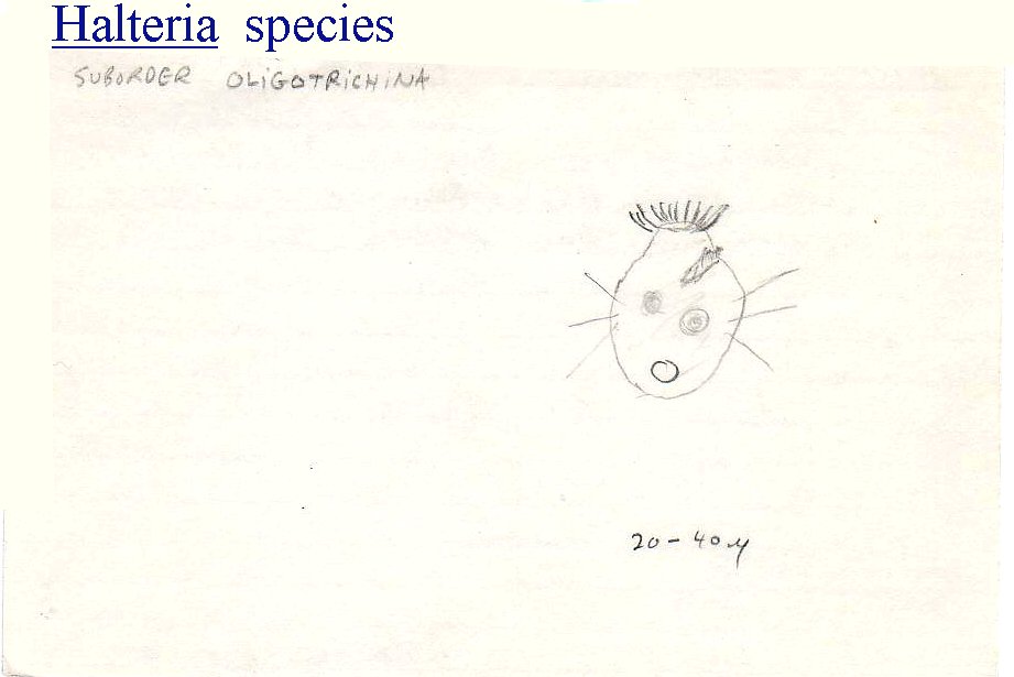
Halteria species:

Characteristic "bouncing ball" motility. Also will rotate in a stationary position. Seen in Kline waterfall stagnating sample 2/84, 5/86. Mud puddle algae 6/84. Delta Slough 8/84. Delta Park Lake 8/84, 6/85. Delta Heron pond 10/84. Klineline 8/85, 4/88. Ridgefield slough 10/85. Long Beach marsh 10/86. Salmon Creek 5/97.
Holophrya species:

Long Beach marsh 4/86. Cilia uniformly long. Cytostome small. Contractile vacoule posterior.
Holosticha species:

2/1987 in old sample from Kline waterfall.
Homalozoon species:

Similar to Spathidium but larger, more elongate, no cilia on one side, and many contractile vacoules in a row. Seen 3/86 from Klne pond algae, 1/87 among oscillatoria algae. Not listed in Jahn, See Kudo pg 839
Hypotrichidium species:

5/1997 many seen in one day old sample from Vancouver Lake backwater plants. Binary fission and conjugation observed. Observed it ingesting a Coleps
Lacrymaria olor The "Swan Animalcule":

One of the most entertaining protozoa to watch! The "neck" is extremely extensible, flexible and active. Fresh or salt water. Seen 3/85 in old Duckweed debris, Kline waterfall 3/85, 2/87, Klineline 8/85, 7/86, Kline pond 7/87, 10/87, 10/88. I have also seen a small Lacrymaria species that measures only 40 microns contracted. The first sighting of this was inside of a dead dinoflagellate shell. Second sighting 2/87 was in marsh algae.
Lembadion species:

10/87, 10/88, 3/92, 1/93, 2/97 from Kline pond. The 1/93 sighting was from algae incubated after collection from under pond ice. Observed it try to capture large flagellate unsuccessfully. 1988 sighting was in a diatom bloom and observed ingested diatom.
Litonotus fasciola:

Active probing ciliate. Cytostome is long slit. Posterior contractile vacoule. Two macronuclei. Seen at Kline waterfall 8/84, 3/85, and 2/87, also Klinepond 10/87
Loxodes species:

8/97 Loomis Lake. Flattened cell.
Loxophyllum meleagris:

Form and size variable, very flexible. Shaped like a broad leaf. Trichocysts in wart like protrubences. Long canal to contractile vacoule. Feeds mainly on rotifers. COOL!
Klineline algae 8/85, 10/87, Loomis lake 8/1997.
Mesodinium pulex:

Marine and fresh water. Marine form seen at LongBeach 8/84 was green. Fresh water sightings were at Klineline 5/86, 4/88. Motility is active, jerky, and bouncy like Halteria.
Monochilium frontatum:

Active bacterial and diatom feeder. Kudo mentions it feeds on algae. Very observable trichocysts. Resembles Frontonia except smaller and oral area more to side. Observed trichocyst discharge. 5/86 Ditch water.
Mycterothrix erlangeri:

Gelatinous lorica, swims backwards. Rotates in lorica slowly and presses particles with side cilia into gelatinous matrix, thereby building tubular lorica. Kudo mentions zoochlorellae but the 9/86 observation did not demonstrate this. Perhaps this was because it was from a temporary mud puddle. It was among mud and oscillatoria.
Nassula aurea:

Active and colorful ciliate. Colored by partially digested algae. Food vacoules are green, yellow, and red. Was observed to stop to ingest large food particle. The 6/88 sighting had many, watched one ingest chain of blue-green algae. Algae protruding from cyrtos.
7/84 Mud puddle with algae
6/88 Tannin colored frog pond water.
2/1997 Red pigmented Klineline sample.
Opisthonecta-henneguyi:

Formerly called
Teletrochidium At first observation this looked like the teletroch stage of a large Vorticella species, but it persisted for days in large numbers in an established Euglena loaded sample from a green puddle in a horse stall. 3/97. Methyl-cellulose plus india ink demonstrated the ciliated cytopharynx quite well. Appears to eat Euglena, also ingested india ink. But only ingested yeast rarely. This is odd since yeast are intermediate in size. Cyst forms found.
This species was found again at the same site in 3/2012. See the
YouTube video
Paramecium aurelia:

Smaller than P. caudatum and P. multimicronucleatum. Rounded posteriorly. Two vesicular micronuclei. One large macronucleus. Trichocyst discharge observed. Cells contain polarizing crystals. The 2/92 sample was cultured into 4 clone cultures. After 7 days cultures showed 10 to 50% cells in autogamy.
Delta Lake 9/84, 5/85, 6/85. Ditch 5/86. Aquarium 5/86. Kline waterfall 3/87, 7/87. Sidewalk algae puddle at Klineline Park 2/1992.
Paramecium bursaria :

Foot shaped Paramecium with symbiotic zoochlorellae. Two contractile vacoules. Good organism for culture in 1/2 strength hay infusion - supply light for symbiotic algae. Cultured organisms when stationary will respond to sudden light increase by spinning briefly towards the convex side of the cell.
Click here to see Youtube video of the organism, including the response to sudden light bursts
Click here to see nucleus stained with Methyl-Green
Delta Park Slough 5/84. Kline Waterfall 8/84. Duckweed Ditch 2/85 this same sample still had P. bursaria after 4 months. Klineline 8/85 Many in Cat-tail pond vegetation debris. Tiny spring 10/85 Delta Lake 6/86. 9/87, 2/92, and 4/94 in Kline pond. 4/92 Klineline lake and again on 1/93 under ice sample incubated 5 days.
Paramecium caudatum :

Cultured 5/84 - still viable on 11/84. Posterior end more pointed than other species. Two contractile vacoules. Usually have more food vacoules than the sketch shows.
4/84 and 3/87 Stagnating Kline waterfall sample. Klineline old algae 8/85 & 5/86. Ditch 5/86
Paramecium trichium (putrinum) :

Rotates to left. Large numbers found in diatom scum in the Ocean Park stream 4/92. Cultured these using P.aurelia media and techniques. Had to flatten stained cells to see micronuclei. Responds to increased light similar to P. bursaria. Cells contain polarizing crystals similar to P. aurelia. Contractile vacoules are fed by vesicles.
Pleuronema species:

Small ciliate, mostly stationary with rotation. Occasional jumpy motility. Slight swelling near oral area distinguishes Pleuronema from Cyclidium species. Darting motion is almost always associated with tactile response, even passing motile bacteria will set it off.
Podophrya fixa:

A common suctorean. 5/84 very old sample from Kline waterfall. 2/85 Duckweed ditch.
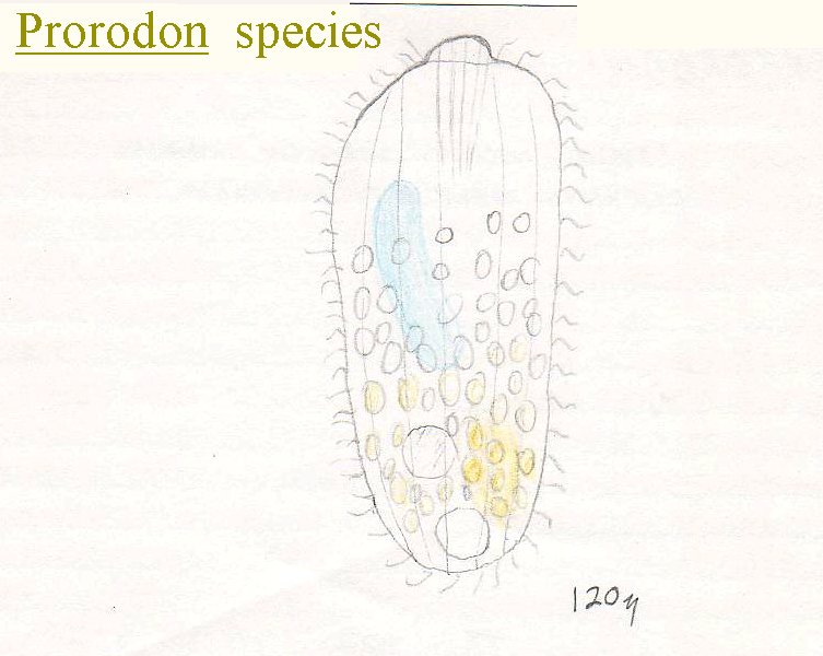
Prorodon species:

Massive nucleus seen on staining. Trichites deep into ectoplasm. 3/92 seen in Whipple Creek ditch sample that Joel collected.
Pyxicola species:

Brown lorica and operculum that closes lorica when organism contracts. Similar to Vaginicola and Cothurnia except for operculum. Also Vaginicola has no pedicel. All three genera found at same location: 9/87 Kline pond algae. Also seen 7/90 at same site.
Pyxidium vernale :

Simple stalk. No lorica. 9/84 Old sample from Delta Lake.
Rhabdostyla to put in
Solenophrya species:

Suctorea: Body not filling lorica. Tentacles in fascicles. Contractile Vacoule fills up to 1/4 of cell volume when full. 2/92 Algae bloom infusion.
Spathidium spathula:

Active voracious carnivorous ciliate. Ingests small ciliates, seems to ignore amoebas. Actively pursues Chilodonella, Colpidia, etc. but ingestions are not frequent. 9/84 and 5/85 Delta Lake mud. 2/85 mud at waterfall site.
Spirostomum intermedium:

Large, plastic, large contractile vacoule, nucleus not seen without stain.
Spirosomum minus:

This specimen had a pinkish hue not mentioned in the references. 10/85 old jar Klineline algae sample.
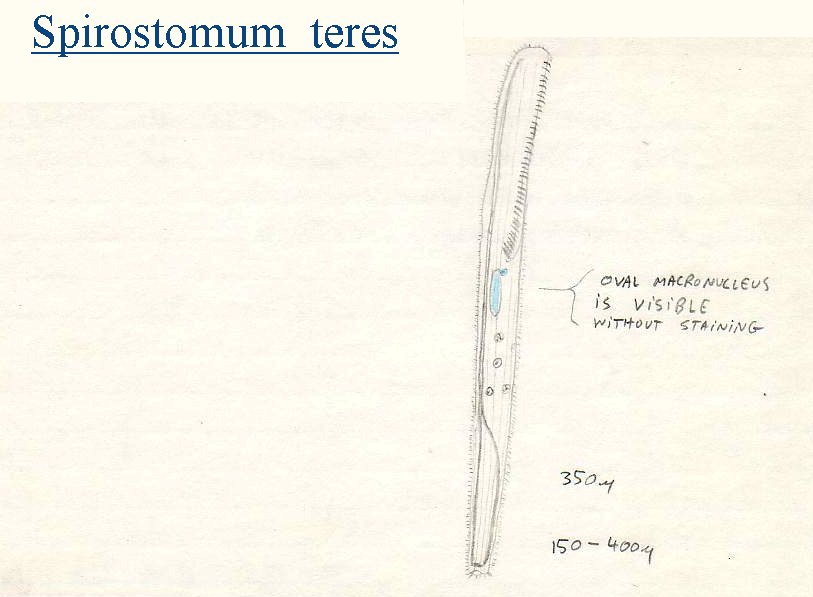
Spirostomum teres:

Distinguished from othr species by one oval macronucleus. Very contractile cell via myonemes.
Stentor amethystinus:

Blue-green pigment. Habitually pyriform (contracted - free swimming) 10/1987 Klineline oscillatoria sample.
Stentor coeruleus:

Brilliant blue-green pigment called stentorin is located in pellicle in inter-striation granules. Environmental changes can cause shedding of this pellicle. Very contractile. Klineline 8/85, 10/86. Delta lake sludge 6/86.
Stentor igneus:

Red pigment. Oval macronucleus. Ciliation uniform. Attachment organelles very obvious on 4/88 sighting - ditch algae bloom.
Stentor polymorphus:

Large. Green with symbiotic algae. Kline-pond 2/92.
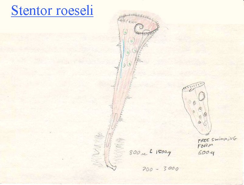
Stentor roeseli:

Faint rose color, but is not S.igneus due to longer cilia groups. Appeared to have gelatinous tube at base. Nucleus long band form. Many sightings at Klineline-pond and Klineline-lake. Also seen in aquarium and Delta Lake mud.
Stichotricha species:

Lives in tube of plant debris. Observed it leave its tube and begin to build a second tube. It took 5-10 minutes to build the first 1/3 of the tube starting from the back end. Found another tube that was 600 microns long. Tube makes observation difficult. Seen in puddles 5/86, 2/92. Kline-pond 10/87 Klineline 4/88. Many in ditch algae bloom 4/88.
Strobilidium gyrans:

Long anterior cilia. Rotates rapidly as either free swimming or stationary rotation due to invisible "mucous" strand. Many ingested diatoms seen in 2/85 sample. Standing fresh water oligotrich ciliate. Swimming motion is similar to Halteria species.
Strombidinopsis species:

Lorica often absent.
Seen 2/85 in Suavies Island marsh.
Stylonchia mytilis:

Image drawn is dorsal with ventral cirri showing through cell. Behaviour is active stalking. When adoral membranelles contact smaller protozoa the Stylonchia will lunge forward rarely ingesting object. Food appears to be mostly bacterial. For this bacterial feeding the organism will sit stationary and generate currents that draw the food in. Observed one ingest a Paramecium once!
Stylonchia species:

Eight frontal cirri, 5 ventral, 5 anal, 3 caudal, and many marginal cirri.
Tetrahymena species:

Active in stagnating samples, bacterial feeder, @ cilia lines to oral ciliature/membranelles.
Tokophrya quadripartita:

Observed in Sewage treatment plant sample/ PSU Protozoology course 4/1997.
Trachelocerca species:

MARINE: 7/97 Ocean Park beach sand and plankton scum. Pellicular striae are not spiral. No "neck" therefore not Chaenae. No ring furrow so not Lacrymaria. There are many Trachelocerca species in salt water environments.
Urocentrum turbo:

Characteristic rotation swimming and off center "tail" cilia tuft make this an easily recognized species. Will rotate stationary with a "mucous" string attachment or swims forward very rapidly. Eight collecting canals connect to posterior contractile vacoule.
Uroleptus longicaudus:

2/1987 old sample from Kline waterfall. Drawing is of ventral side.
Urostyla species:

Active hypotrich...(aren't they all !?) 8/85 Klineline old algae sample.
Urosoma caudata:

Active hypotrich...(aren't they all !?) Long posterior end is clue to this ID. 9/84 Drying river bed.
Urotricha species:

Small ciliate with anterior cytostome, posterior contractile vacoule and long caudal cili (is "cili" singular for cilia ?). Seen twice from marshes and once from a stagnant sample from Klineline.
Vaginicola species:

Feeds like Vorticella. Retracts on stimulus. One contractile vacoule. Lorica can be clear to yellow to brown. See also Cothurnia and Pyxicola species.
Vorticella campanula:

Observed sexual and asexual reproduction. Large species with wide oral area. Many internal refractile granules.
Vorticella convallaria:

No refractile granules and narrower oral area than V. campanula.
Vorticella microstoma:

Narrow oral area hence the species name.
HELP me ID this Suctorean..:

Suctorean with a lorica: I could not find this Suctorean in the literature I have available. If anyone knows what this is please Email me at brodwcjj@integrity.com Thanks.


